Friday, May 19, 2006
Female Uretra Play Vids
We 2A Liceo Tecnologico dell'ITCG F. Fontana di Rovereto, Trento.
The work you are proposing about the deepening of Sciences done during the 2005-06 school year and that had as its object, the microscope .
One tool that has literally changed the way science and history with a spick and that is intertwined in some way, even with Felice Fontana, the person who has been called the school and who has made a fundamental contribution to the development of cell theory.
This was a group effort that lasted a few months and wanted to use a somewhat different kind of "poster" to introduce him here so we chose him: THE BLOG !!!!!!
good read ...
Gratis Bilder Mom Feet
cell theory is one if not "the" fundamental principle of biology. The story of his birth is extremely fascinating. We wanted to go in his milestones, for better appreciate its value but also to understand the ideas and theories not only of scientists of the time as Fontana, Hooke, Schleiden and Schwann that have contributed to this theory, but also to know theories that wants to be antagonistic to it.
The theory is so 'summarized as follows:
- All living organisms are made up of one or more cells
- The chemical reactions of a living organism, including the mechanisms of release of energy and biosynthetic reactions take place within cells;
- cells originate from other cells;
- The cells contain the hereditary information bodies to which they belong, and this information passed from the mother cell to daughter cell.
below illustrate the most important steps that have contributed to the formulation of this fundamental theory: In
seventeenth century. Robert Hooke (1635-1702), a great mathematician, physicist, naturalist and astronomy using a microscope of his invention, he noticed that the cork and other plant tissues were formed by small cavities, separated by walls called "cells" or "little room". In 1838
CE rather Matthias J. Schleiden (1804-1881), German botanist, concluded that all plant tissues are built from sets of organized cells.
In 1839 AD Theodor Schwann (1810-1882), German zoologist, extended Schleiden's observations to animal tissues and proposed a cellular basis common to all living beings. In 1840
is made an introduction of the cell theory. Included the results of experiments and research of many scientists. Among the precursors of this theory was also Felice Fontana.
Felice Fontana (1730-1805) was a scientist in Trentino. During his studies on the skin of eels, was one of the first without the help of microscopes, saw CELL. And the first one who noticed the nucleus of an adult animal cell. Here is his remark made at Naples in 1787, "I had curious to examine the skin of eels gluten, my stools they bring many different thicknesses, and found, after a bit 'diluted, and taken to have a tiny amount, ch' He seemed composed of uniform and irregular blisters, filled with tiny corpiccioli, spheroidal "
In 1858 AD Rudolf Virchow (1821-1902), pathologist, said that the cells may originate only from cells background. When a cell exists, there must be a pre-existing cells, just like an animal arises only from an animal and arises only from a planet a planet.
In 1859 AD new conventions emerged from the evolutionary theory of Charles Darwin: "There is a strong continuity between existing cells and cells that first appeared on Earth"
Microscope
By 1800 the microscope was fallen out of favor for several ragioni.I compound microscopes used in the 18th century, with their systems of lenses with numerical apertures and incorrect available, suffered a high degree of aberration (distortion) chromatic and spherical. With the magnification used (Up to 500 times) images were often confused, and were the dominant optical false, so that these observers largely completed their observations with the fantasies and the microscope had come to serve primarily as a means of entertainment.
Felice Fontana was well aware of the problems of microscopy, and discussed the "tiny mistake and the consequences deduced from microscopic observations. In the Treaty on the venom of the viper, 1787, he wrote: "... A simple bare observation of the whole, may not deserve full confidence, because we assume that there is a necessary and exclusive relationship between the image represented by the microscope real external object ... "
spontaneous generation
Until the middle of seventeenth century it was common belief that God created humans and other higher organisms, and amphibians, worms, the insects would be generated spontaneously from mud. This belief has its origins far removed not only in terms of time but also space. In China it was thought that the bamboo amphibians were born, for the Greek philosophers, life was contained in the material itself and from it emerged spontaneously when the conditions were made favorevoli.La theory of spontaneous generation step through the Middle Ages and the Renaissance and was supported by great thinkers like Newton, Descartes. From the mid-seventeenth
began, however, the first experiments in support of the cell theory. In 1668, the Tuscan FRANCESCO REDI illustrated a series of experiments that were supposed to disprove the generation spontanea.Egli put the veal and fish in some hermetically sealed containers that, leaving others open. After a bit 'of time could note the presence of maggots on rotting meat inside the open containers in which the flies came and went freely, while there was no trace of the sealed containers. Isolated in a separate container and saw that these worms after various transformation, becoming the adult flies. Redi this experiment had proved beyond doubt that the larvae do not come from rotting flesh.
More or less the same time, it did make important discoveries, the naturalist Anton van Leeuwenhoek . It was the first to discover the microscope or to use magnifying glasses, was the first to see and describe bacteria. He was a cloth merchant who lived in the Netherlands and used the lens to see the quality of the materials. In the 1668 trip to England to see English fabrics, found slower more powerful than he had. Back in Holland he developed and tested the lens looking at every thing he had before, until you see the microbes. He made numerous microscopes in silver and gold. His best lenses were 300-500 times larger, allowing him to see protozoa and large batteri.La generation was then transferred to these cards. In fact, the rotting flesh in the long run also gave rise to micro-organisms in closed vessels.
gave an answer to the latter theory, the Italian naturalist Lazzaro SPALLANZZANI , who repeated the experiment, but boiled and hermetically sealed part of the flesh; result, this time even microorganisms could svilupparsi.La dispute continued for many years and finally ended in the mid-nineteenth century when the French biologist Louis Pasteur devised an experiment that would put an end to dibattito.Pasteur personally constructed of glass containers with a long curved neck (swan-neck flasks), which was placed inside the nutrient solution which was boiled for an hour letting the steam came out freely from the orifice end of the curved neck. Off the heat, because of depression caused by the heating, air contaminated by bacteria and other microorganisms. Those in contact with the liquid that were still boiling inside, were killed. After several months, the infusion was still stored in clear demonstration that there were no seeds of any kind, while the outer section of the neck you could see the presence of dust and microorganisms entered the opening terminale.In more clearly showed that the boiling liquid, he had not killed the "active" because it was enough to snap off the neck twisted by placing the liquid in contact with the nutritional ' air, after a few hours would be numb to the presence of spores and germs. What
of spontaneous generation is not the only case in which the scientific community has interpreted natural phenomena on the basis of the existence of substance which is then frantically went to ricerca.L 'air has long been populated by "Vis Vitalis, but also from phlogiston, heat, ether. The conviction we have today that these theories were not true and justified, we only thanks to a rigorous theoretical and experimental research and also thanks to the invention of the microscope and its continuous improvement.
Errore You Are Sending Spam
The microscope was a slow process and sometimes random findings, by scientists but also of simple artisans. It is thanks to this series of discoveries and small steps that the optical microscope was born and grew.
property of lenses and mirrors were known since antiquity, but were never studied sistematico.Gli same problems that blocked the development of the telescope, also prevented the advancement of microscopy, optics linked to that. Higher members of this advancement are Galileo, Cornelius Drebbel, Anthony van Leeuwenhoeck, Robert Hooke . The theory of optical microscope
progressed rapidly, but so did the practice: in fact for many centuries the most used tool was the simple microscope. The latter is, for example, that of van Leeuwenhoeck or with a single lens system, while of the dialed is an example of what Hooke was not competitive for a long time because of the difficulty of construction and defects of the lenses. Only the improvements introduced by the microscope Giovanni Battista Amici and Ernst Abbe situazione.I allowed to change the technological advances brought on, in modern times to X-ray microscopy and electronic ones.
chronicles report that the microscope was founded in 1590 in a Dutch lens maker's shop, just as the telescope twenty years later. The development of the telescope however, is quick and overwhelming, while his brother remains for more than fifty years forgotten along with other quirky objects that filled his time. Why the difference? The telescope had revolutionized astronomy, a discipline from ancient traditions, and so had become the symbol of the new science that within a few decades would have wiped out the ancient tradition of Aristotle. Instead of a microscopic reality not imagine even the existence, let alone was the focus of philosophical clashes. The first models did not even aroused great interest, they simply show the ordinary world of everyday life just a little 'more magnified. Yet the path of the microscope was already sealed. But it takes years to slow and focus be improved, but eventually the microscope awakens the interest of scientists.
The work confirms the reputation of this tool is the "Micrographia" Hooke, published in 1665. It is not only the precision of observations that determine the success of the book, since the use of beautiful and detailed pictorial. Scientists are impressed by that monstrous and exotic world, full of flies and lice from the compound eyes hairy legs! The "Micrographia" is immediately adopted by the European universities, and remains in use for well over a century. As if to say that the book on which you are studying may be the 1900 or so!
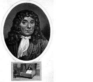
Leeuwenhoeck Antony van (1632-1723)
The story takes an unexpected turn when the microscope of the cross Leeuwenhoeck , a curious figure of amateur science. Anthony van Leeuwenhoeck was a wealthy cloth merchant, the equally incredible dexterity and boundless curiosity. It is not a scientist and does not understand the Latin (which were written most of the scientific books of the time) but not lose heart. Home study achieves unsurpassed skill in cutting lenses, yielding magnifications much higher than those of other microscopists. His boundless curiosity does the rest, bringing it to examine not only insects but also parts of crystals, grains of pepper, seeds, blood, milk, crushed rocks and anything that happens under his hand!
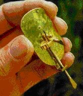
The structure of the microscope used by Leeuwenhoeck in an exact replica modern
Simplicity and dimensions of this instrument is extraordinary.
summer of 1674 Leeuwenhoeck is pass by a pond and decides to submit to its slow even that water greenish and smelly. Imagine what was his surprise when he discovered a huge amount of tiny little creatures, much smaller than any insect or worm then known, and fluttered frantically! A new world opened before him, swarming with strange forms of animals. And all this in an ordinary drop of water! Leeuwenhoeck public comment. They are the first work that describes protozoa and bacteria, and have a huge resonance.
The microscope then becomes, suddenly, very popular: it is broken out fashion stagnant water!
Hosts of reputable scholars and amateurs leave the investigation of animal and vegetable tissues, which may have provided more interesting, and launch into passionate study in miniature of those worms that seem to be everywhere (Leeuwenhoeck believes himself to see them even in salt or in the dust!). From this research, mostly inspired by simple curiosity, you will slowly to a rigorous and systematic use of the microscope. Thanks to this extraordinary instrument
cells, observed for the first time by Hooke, will be recognized in the first half of the nineteenth century as the key elements of living matter. It will be one of the greatest revolutions in the history of science, and will mark the birth of microbiology.
also strides that medical research has done over the past 100-150 years, with everything that follows for our health, would have been unthinkable without the microscope. That will evolve and become even a scanning electron microscope and atomic then, increasing its size to "see" even the atoms. Today, the microscope is an invaluable tool in the laboratory, and became the very symbol of scientific research.
Think about it: what are the symbols of science? The atom surrounded by electrons, Einstein, the computer and the microscope. Not the telescope. Here, after five centuries, the tiny microscope has taken its well deserved revenge on his brother, the telescope!
Sources: Enkarta, Encyclopedia of the Republic.
Authors: Matthew Mayr, Kevin Zomer, Serena Martini.
Thursday, May 18, 2006
Free One Nite In Paris
Introduction:
The electron microscope is based essentially on the same principles as the optical, but it does provide greater magnification, but the downside is that the use of techniques and equipment preparations and interpretation are more complex and expensive.

Instead of the light source used in the optical microscope, the light is replaced by a accelerated electron beam in a vacuum, rather than using the lenses are used for electric and magnetic fields that have a convergent effect on the electrons. Since the electrons are associated with a wavelength (the wavelength is the distance between two consecutive crests of a sine wave)
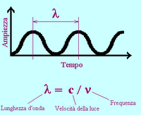 much smaller than that of the visible spectrum this leads to an increase in resolving power up to about 10 Å. Even if we can get to these very high resolutions in general, however, it uses a 200 times greater than that of MOC (Compound microscope) for technical deficiencies that prevent the exploit the theoretical possibilities.
 remember also that electron microscopes can see the black and white samples that are only after "colored."
A curiosity is that the electric microscope, as the test material is placed in a vacuum, it must first be dehydrated, which precludes the use of electron microscope preparations on living tissue sections also must be very thin to allow electrons which have a very low penetration power, to pass.
The scanning electron microscope (SEM):
functionality of this microscope is analogous to a closed-circuit television cameras. The sample surface is hit and pierced by an electron beam that scans the surface by means of a deflector roll, moving like on a television screen (the number of lines per picture is around 100 to 2,000). The secondary electrons generated point by point from the surface are collected by a collector electrode at 200 V.
The electrode produces an electric signal that modulates the electron beam on the screen, in sync with the primary beam path: the screen image is formed with a large depth of focus. The only limitation of this tool is that the sample should be analyzed under high vacuum: this was developed for the environmental scanning electron microscope, which is no longer bound by this limit, and is capable of analyzing samples of organic material while maintaining the conditions of temperature, pressure and humidity.
The use of electrons instead of visible light has a number of significant advantages for performance of the instrument.
- The minimum wavelength of visible light, in fact, is about 400 nm (400 millionths of a millimeter), while the wavelength associated with the electron in these tools may be of only 0.05 nm.
- The resolving power of electron microscope is therefore significantly greater than that of optical microscopes: we come to perceive details of a few tenths of nm . (1 nm = 10 -6 mm)
look at an eye of an insect with two types of microscopes:
light microscopy
 electron microscope

http://ion.asu.edu/live42_wasp/live42_wasp_thumb.htm
The transmission electron microscopy (TEM) electron microscope
In transmission using an electron beam as the X-ray microscope and scanning electric microscope. The electrons are collected and concentrated by a magnetic spot on the sample capacitor. In the test material occurs diffraction, which reach the goal, which is also strictly magnetic, and then to a projector which transforms and sends the image to fit a screen fluorescente.Di result condenser lens and lens are not made but by magnetic fields, which serve to deflect the trajectories of electrons moving towards the axis. The source is very often made of a tungsten filament, whose potential is negative and remained between 30 and 10 kV.
varying the intensity of the current in the circuit of the capacitor varies not only the intensity but also the convergence electron beam incident. By varying the
instead of our projector sets the magnification, while varying the lens improves the clarity.
Magnification is variable and depends mainly on the intensity of current through the coils of the projector. They can also be provided with an electron gun from a MV that give a resolving power between 2 and 5 and magnification up to 106 diameters.
Authors: distort, LC, Dosso
Saturday, May 13, 2006
Example Welcomw Wddress

optical microscope
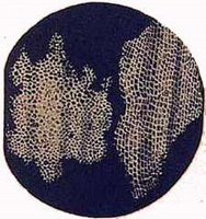 Observation of a thin section of cork
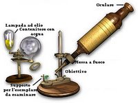 components of an optical microscope ancient
 microscopes are instruments used to produce enlarged visual images of objects too small to be observed with the naked eye .
It must fulfill three basic functions: 1
produce a magnified image of the preparation,
2 separate 3
details make this visible to the eye.
The full name of the microscope is " optical microscope", which is useful to distinguish it from simple microscope known as a magnifying glass. Essentially
the optical microscope consists of two converging lenses and convex.
These two lenses are in 'target and in' eye, placed at each end of a tube called the body tube of the microscope.
The goal, placed near the object to be observed real produces an image, magnified and inverted, which in turn is viewed through the eyepiece, it receives this image in its own fire and turns in the final image, which is virtual, inverted and larger than the real one.
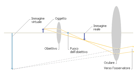
To understand what is happening is also important to understand what happens when our eyes see the image.
.
Authors: Massimo Gaggia, Giori Fabrizio, Kristian Sartori
Sources : http://micro.magnet.fsu.edu ; http://it.encarta.msn.com ;
Tiffany Granath Y Julie Ashton
This report was made to publicize what we have achieved during the hours spent in the laboratory science, during which we made the simple experience of microscopy that now we will describe:
1) Experience on " Osmosis "
- Osmosis is the movement that make the water molecules in crossing a semipermeable membrane ( = membrane that allows passage only to certain substances of very small ), this process takes place between a solution that has a solution with higher concentration of solutes and one with a child.
- Phases of the experiment:
1. We cut a piece of onion skin (very thin, otherwise you will not be able to see the purpose of the experiment);
2. We put on a glass slide preparation, we have paid a couple of drops of water, we put on the table for objects and we have adjusted, over the light source, means of travel wheels;
3. Looking through the eyepiece of the preparation we note that its color is the pink / red, his features are clearly visible and the cell fills the latter;
4. add a few drops of saline (NaCl ) and we note that the cell does not ricropre plus all the space on the first but is detached from its cell wall, as if it was wrinkled;
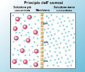 The movement of particles in the osmotic process
2) Experience on " Chloroplasts: Chloroplasts are
-cellular organelles that contain chlorophyll and photosynthetic processes occurring within them ( are to transform ' light energy into chemical energy ).
- Experiment:
- The experiment consists in observing a leaf of elodea ( Elodia canadensis, an aquatic plant commonly called Plague of water because it also infests the waters of the rivers ).
1. prepare the specimen for observation under a microscope;
2. The product comes with small organelles arranged at random within the cell are green;
3. add saline solution and observe that the chloroplasts are joined in a long chain inside the cell;
4. If we increase the magnification of the microscope we can see the small bodies that move randomly within the cell;
 elodea cells under a microscope
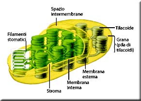 model chloroplast
3) viewing experience of " paramecium "
- The paramecium is an aquatic organism, or stagnant fresh water, unicellular, belongs to the family of protozoa. Their shape is round-oval lashes evenly distributed over the entire surface of the cell, which used to move and to feed mainly of bacteria through a slot called " cytostome " and eliminate waste matter through " citopiglio .
-Experiment:
1. We took a sample of stagnant water from a tank specially prepared to study these organisms;
2. We did a little 'hard to find one, because it moves so quickly and why then you can do with a very high magnification, but in the end we succeeded;
3. During the research we have found other bodies much larger, a 30th of times, but slower, their movement resembles the movement that makes a "snail" is crushed and then rieste;
here is a picture of a paramecium:

4) Experience on " amyloplasts "
- belong to the group of Pastida, relatively large organelle of plant cells that take part in photosynthesis, its job is to store starch (carbohydrate = stored as reserve materials ).
-Experiment:
1. Prepare the prepared by cutting a banana and proceeding as explained above;
2. In the banana amyloplasts are all clustered at the center of the cell
3. we note that increasing the magnification of the nuances, he discovers that the rings are growth;
4. add the dye in the preparation and it colors everything we see that even in the preparation of the cell, it does not allow us to see anything else because we can not distinguish the good features of the cell and amyloplasts;
Authors: Iacopo Smaniotto, Tomasoni Emanuele, Marco Pied.
Sources taken:
- the Encyclopedia Repubbliaca;
- Books: "Invitation to Biology"
- Notes taken during the hours of lab sciences,
- In pictures: Internet;
Friday, May 12, 2006
How To Cutchoridar Pajama
Some basic rules on the use

2) The distance of the eyepieces should be the distance between the eyes of the observer. Then adjust the distance between the eyepieces to see a 'single fused image.

3) the appropriate position (with the part to be tested facing the lens) the slide on the mechanical stage (the object must be observed to transmit light otherwise you do not see anything).

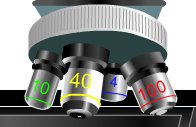
5) Select the objectives of the magnification of the instrument, starting lower and then gradually progressing up to the greater.
6) Put "focus" the sample by turning the appropriate knob on the side of the larger microscope.

7) Observe carefully (if necessary by increasing the beam with the diaphragm).
Sources: http://www.nsgs.it/microscopioa.html
