electron microscopes
Introduction:
The electron microscope is based essentially on the same principles as the optical, but it does provide greater magnification, but the downside is that the use of techniques and equipment preparations and interpretation are more complex and expensive.
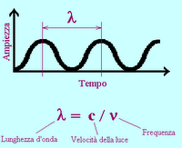
Instead of the light source used in the optical microscope, the light is replaced by a accelerated electron beam in a vacuum, rather than using the lenses are used for electric and magnetic fields that have a convergent effect on the electrons. Since the electrons are associated with a wavelength (the wavelength is the distance between two consecutive crests of a sine wave) much smaller than that of the visible spectrum this leads to an increase in resolving power up to about 10 Å. Even if we can get to these very high resolutions in general, however, it uses a 200 times greater than that of MOC (Compound microscope) for technical deficiencies that prevent the exploit the theoretical possibilities.
remember also that electron microscopes can see the black and white samples that are only after "colored."
A curiosity is that the electric microscope, as the test material is placed in a vacuum, it must first be dehydrated, which precludes the use of electron microscope preparations on living tissue sections also must be very thin to allow electrons which have a very low penetration power, to pass.
The scanning electron microscope (SEM):
functionality of this microscope is analogous to a closed-circuit television cameras. The sample surface is hit and pierced by an electron beam that scans the surface by means of a deflector roll, moving like on a television screen (the number of lines per picture is around 100 to 2,000). The secondary electrons generated point by point from the surface are collected by a collector electrode at 200 V.
The electrode produces an electric signal that modulates the electron beam on the screen, in sync with the primary beam path: the screen image is formed with a large depth of focus. The only limitation of this tool is that the sample should be analyzed under high vacuum: this was developed for the environmental scanning electron microscope, which is no longer bound by this limit, and is capable of analyzing samples of organic material while maintaining the conditions of temperature, pressure and humidity.
The use of electrons instead of visible light has a number of significant advantages for performance of the instrument.
- The minimum wavelength of visible light, in fact, is about 400 nm (400 millionths of a millimeter), while the wavelength associated with the electron in these tools may be of only 0.05 nm.
- The resolving power of electron microscope is therefore significantly greater than that of optical microscopes: we come to perceive details of a few tenths of nm . (1 nm = 10 -6 mm)
look at an eye of an insect with two types of microscopes:
light microscopy
Introduction:
The electron microscope is based essentially on the same principles as the optical, but it does provide greater magnification, but the downside is that the use of techniques and equipment preparations and interpretation are more complex and expensive.

Instead of the light source used in the optical microscope, the light is replaced by a accelerated electron beam in a vacuum, rather than using the lenses are used for electric and magnetic fields that have a convergent effect on the electrons. Since the electrons are associated with a wavelength (the wavelength is the distance between two consecutive crests of a sine wave)
 much smaller than that of the visible spectrum this leads to an increase in resolving power up to about 10 Å. Even if we can get to these very high resolutions in general, however, it uses a 200 times greater than that of MOC (Compound microscope) for technical deficiencies that prevent the exploit the theoretical possibilities.
 remember also that electron microscopes can see the black and white samples that are only after "colored."
A curiosity is that the electric microscope, as the test material is placed in a vacuum, it must first be dehydrated, which precludes the use of electron microscope preparations on living tissue sections also must be very thin to allow electrons which have a very low penetration power, to pass.
The scanning electron microscope (SEM):
functionality of this microscope is analogous to a closed-circuit television cameras. The sample surface is hit and pierced by an electron beam that scans the surface by means of a deflector roll, moving like on a television screen (the number of lines per picture is around 100 to 2,000). The secondary electrons generated point by point from the surface are collected by a collector electrode at 200 V.
The electrode produces an electric signal that modulates the electron beam on the screen, in sync with the primary beam path: the screen image is formed with a large depth of focus. The only limitation of this tool is that the sample should be analyzed under high vacuum: this was developed for the environmental scanning electron microscope, which is no longer bound by this limit, and is capable of analyzing samples of organic material while maintaining the conditions of temperature, pressure and humidity.
The use of electrons instead of visible light has a number of significant advantages for performance of the instrument.
- The minimum wavelength of visible light, in fact, is about 400 nm (400 millionths of a millimeter), while the wavelength associated with the electron in these tools may be of only 0.05 nm.
- The resolving power of electron microscope is therefore significantly greater than that of optical microscopes: we come to perceive details of a few tenths of nm . (1 nm = 10 -6 mm)
look at an eye of an insect with two types of microscopes:
light microscopy
 electron microscope

http://ion.asu.edu/live42_wasp/live42_wasp_thumb.htm
The transmission electron microscopy (TEM) electron microscope
In transmission using an electron beam as the X-ray microscope and scanning electric microscope. The electrons are collected and concentrated by a magnetic spot on the sample capacitor. In the test material occurs diffraction, which reach the goal, which is also strictly magnetic, and then to a projector which transforms and sends the image to fit a screen fluorescente.Di result condenser lens and lens are not made but by magnetic fields, which serve to deflect the trajectories of electrons moving towards the axis. The source is very often made of a tungsten filament, whose potential is negative and remained between 30 and 10 kV.
varying the intensity of the current in the circuit of the capacitor varies not only the intensity but also the convergence electron beam incident. By varying the
instead of our projector sets the magnification, while varying the lens improves the clarity.
Magnification is variable and depends mainly on the intensity of current through the coils of the projector. They can also be provided with an electron gun from a MV that give a resolving power between 2 and 5 and magnification up to 106 diameters.
Authors: distort, LC, Dosso
0 comments:
Post a Comment