This report was made to publicize what we have achieved during the hours spent in the laboratory science, during which we made the simple experience of microscopy that now we will describe:
1) Experience on " Osmosis "
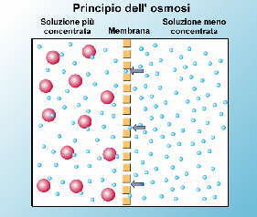
- Osmosis is the movement that make the water molecules in crossing a semipermeable membrane ( = membrane that allows passage only to certain substances of very small ), this process takes place between a solution that has a solution with higher concentration of solutes and one with a child.
- Phases of the experiment:
1. We cut a piece of onion skin (very thin, otherwise you will not be able to see the purpose of the experiment);
2. We put on a glass slide preparation, we have paid a couple of drops of water, we put on the table for objects and we have adjusted, over the light source, means of travel wheels;
3. Looking through the eyepiece of the preparation we note that its color is the pink / red, his features are clearly visible and the cell fills the latter;
4. add a few drops of saline (NaCl ) and we note that the cell does not ricropre plus all the space on the first but is detached from its cell wall, as if it was wrinkled;
 The movement of particles in the osmotic process
2) Experience on " Chloroplasts: Chloroplasts are
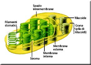
-cellular organelles that contain chlorophyll and photosynthetic processes occurring within them ( are to transform ' light energy into chemical energy ).
- Experiment:
- The experiment consists in observing a leaf of elodea ( Elodia canadensis, an aquatic plant commonly called Plague of water because it also infests the waters of the rivers ).
1. prepare the specimen for observation under a microscope;
2. The product comes with small organelles arranged at random within the cell are green;
3. add saline solution and observe that the chloroplasts are joined in a long chain inside the cell;
4. If we increase the magnification of the microscope we can see the small bodies that move randomly within the cell;
 elodea cells under a microscope
 model chloroplast
3) viewing experience of " paramecium "
- The paramecium is an aquatic organism, or stagnant fresh water, unicellular, belongs to the family of protozoa. Their shape is round-oval lashes evenly distributed over the entire surface of the cell, which used to move and to feed mainly of bacteria through a slot called " cytostome " and eliminate waste matter through " citopiglio .
-Experiment:
1. We took a sample of stagnant water from a tank specially prepared to study these organisms;
2. We did a little 'hard to find one, because it moves so quickly and why then you can do with a very high magnification, but in the end we succeeded;
3. During the research we have found other bodies much larger, a 30th of times, but slower, their movement resembles the movement that makes a "snail" is crushed and then rieste;
here is a picture of a paramecium:

4) Experience on " amyloplasts "
- belong to the group of Pastida, relatively large organelle of plant cells that take part in photosynthesis, its job is to store starch (carbohydrate = stored as reserve materials ).
-Experiment:
1. Prepare the prepared by cutting a banana and proceeding as explained above;
2. In the banana amyloplasts are all clustered at the center of the cell
3. we note that increasing the magnification of the nuances, he discovers that the rings are growth;
4. add the dye in the preparation and it colors everything we see that even in the preparation of the cell, it does not allow us to see anything else because we can not distinguish the good features of the cell and amyloplasts;
Authors: Iacopo Smaniotto, Tomasoni Emanuele, Marco Pied.
Sources taken:
- the Encyclopedia Repubbliaca;
- Books: "Invitation to Biology"
- Notes taken during the hours of lab sciences,
- In pictures: Internet;
0 comments:
Post a Comment